
Screening Center & Imaging Hub
Run fast with industrial-scale screening. See deeper with handcrafted imaging. Seamlessly integrated to achieve our goal.
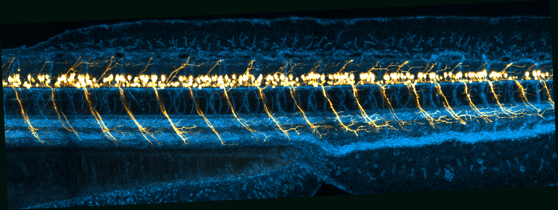
Screening Center
With the coding sequences of vertebrate genomes successfully mapped, the next scientific frontier is clear: establishing the functions of these gene products and decoding how they interact to control cellular activity.
• Systematic Gene Screening: The Screening Centre utilizes tailor-made parallel assays to systematically elucidate gene functions within cells and zebrafish models.
• A Worldwide Unique Resource: In conjunction with the European Zebrafish Resource Center (EZRC), we provide a one-of-a-kind infrastructure. We proudly operate as an open-access user facility, serving both our internal institute and the broader international research community.
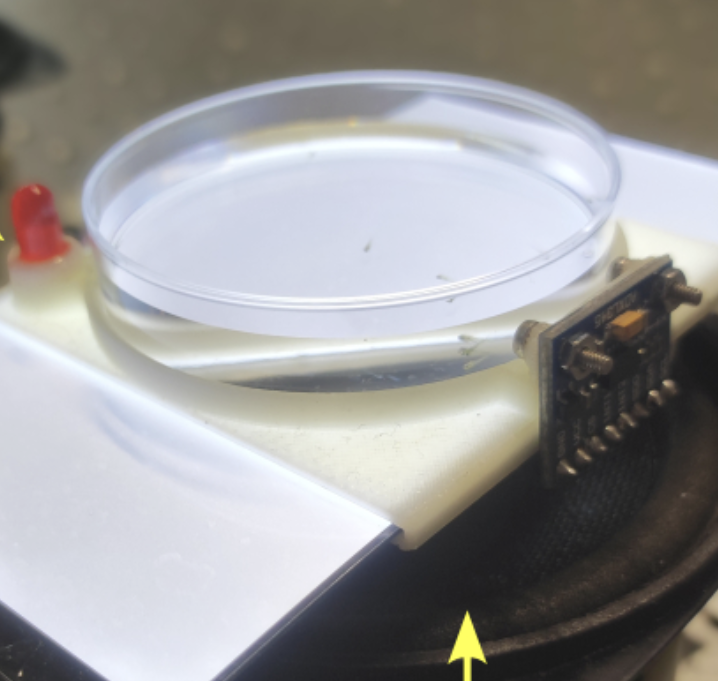
Robust screening system for the evaluation of neuromuscular and general toxicity in zebrafish embryos
Publication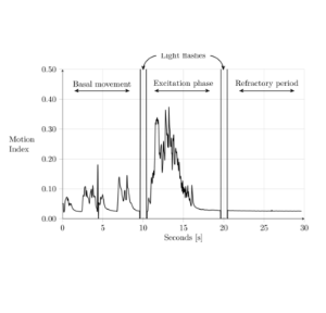
Zebrafish embryos react to the light pulse through activating the nervous system. This platform allows us to screen neuroactive chemicals.
Publication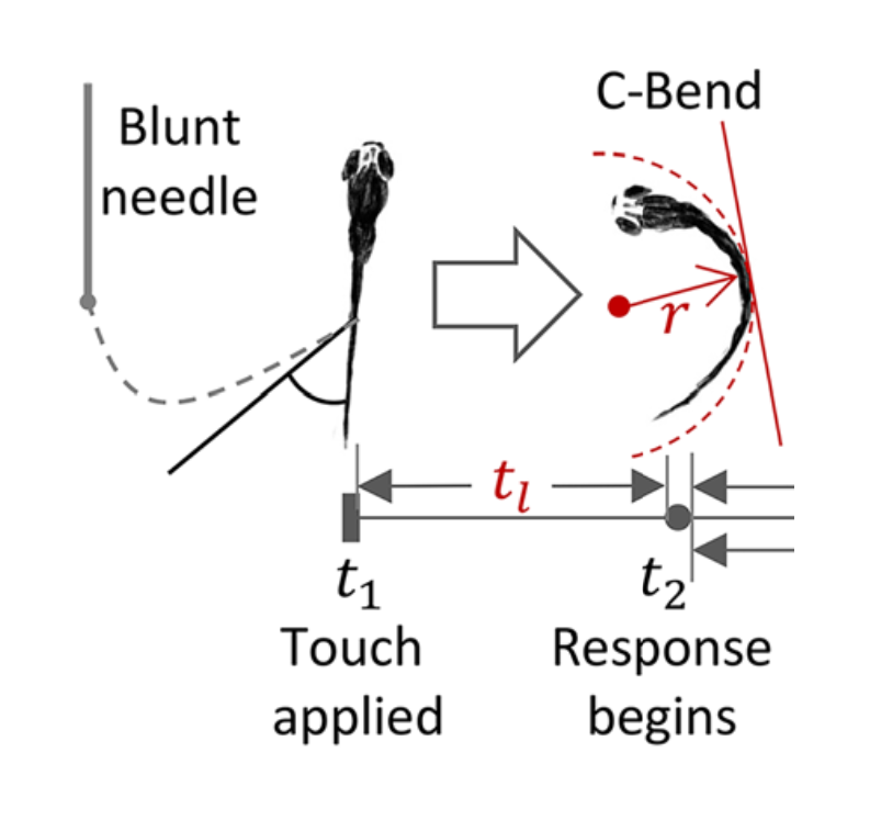
Selected Publications
Hayot, G.; Marcato, D.; Cramer von Clausbruch, C. A.; Pace, G.; Strähle, U.; Colbourne, J. K.; Pylatiuk, C.; Peravali, R.; Weiss, C.; Scholz, S.; Dickmeis, T.
2024. Journal of visualized experiments : JoVE, 203, Art.-Nr.: e66153. doi:10.3791/66153
Cramer von Clausbruch, C.; Weiss, C.; Dickmeis, T.; Peravali, R.; Strähle, U.; Colbourne, J.; Hayot, G.; Scholz, S.; Lopez, R. M.
2023. Naunyn-Schmiedeberg’s Archives of Pharmacology, 396 (S1), S61–62. doi:10.1007/s00210-023-02397-6
Dickmeis, T.; Hayot, G.; Clausbruch, C. C. von; Martinez Lopez, R.; Scholz, S.; Colbourne, J.; Strähle, U.; Weiss, C.; Peravali, R.
2023. Naunyn-Schmiedeberg’s Archives of Pharmacology, 396 (S1), S61
Hansjosten, I.; Takamiya, M.; Rapp, J.; Reiner, L.; Fritsch-Decker, S.; Mattern, D.; Andraschko, S.; Anders, C.; Pace, G.; Dickmeis, T.; Peravali, R.; Rastegar, S.; Strähle, U.; Hsiao, I.-L.; Gilliland, D.; Ojea-Jimenez, I.; Ambrose, S. V. Y.; Belinga-Desaunay-Nault, M.-F. A.; Khan, A. O.; Lynch, I.; Valsami-Jones, E.; Diabaté, S.; Weiss, C.
2022. Environmental science / Nano, 91 (1), 375–392. doi:10.1039/d1en00299f
Gerber, V.; Yang, L.; Takamiya, M.; Ribes, V.; Gourain, V.; Peravali, R.; Stegmaier, J.; Mikut, R.; Reischl, M.; Ferg, M.; Rastegar, S.; Strähle, U.
2019. Development <Cambridge>, 146 (4), dev172510. doi:10.1242/dev.172510
Jarque, S.; Fetter, E.; Veneman, W. J.; Spaink, H. P.; Peravali, R.; Strähle, U.; Scholz, S.
2018. PLoS one, 13 (8), e0203087. doi:10.1371/journal.pone.0203087
Bedell, V.; Buglo, E.; Marcato, D.; Pylatiuk, C.; Mikut, R.; Stegmaier, J.; Scudder, W.; Wray, M.; Züchner, S.; Strähle, U.; Peravali, R.; Dallman, J. E.
2018. Methods in enzymology, 602, 189–209. doi:10.1016/bs.mie.2018.02.004

Fast and Gentle Confocal Super-Resolution for every colour.
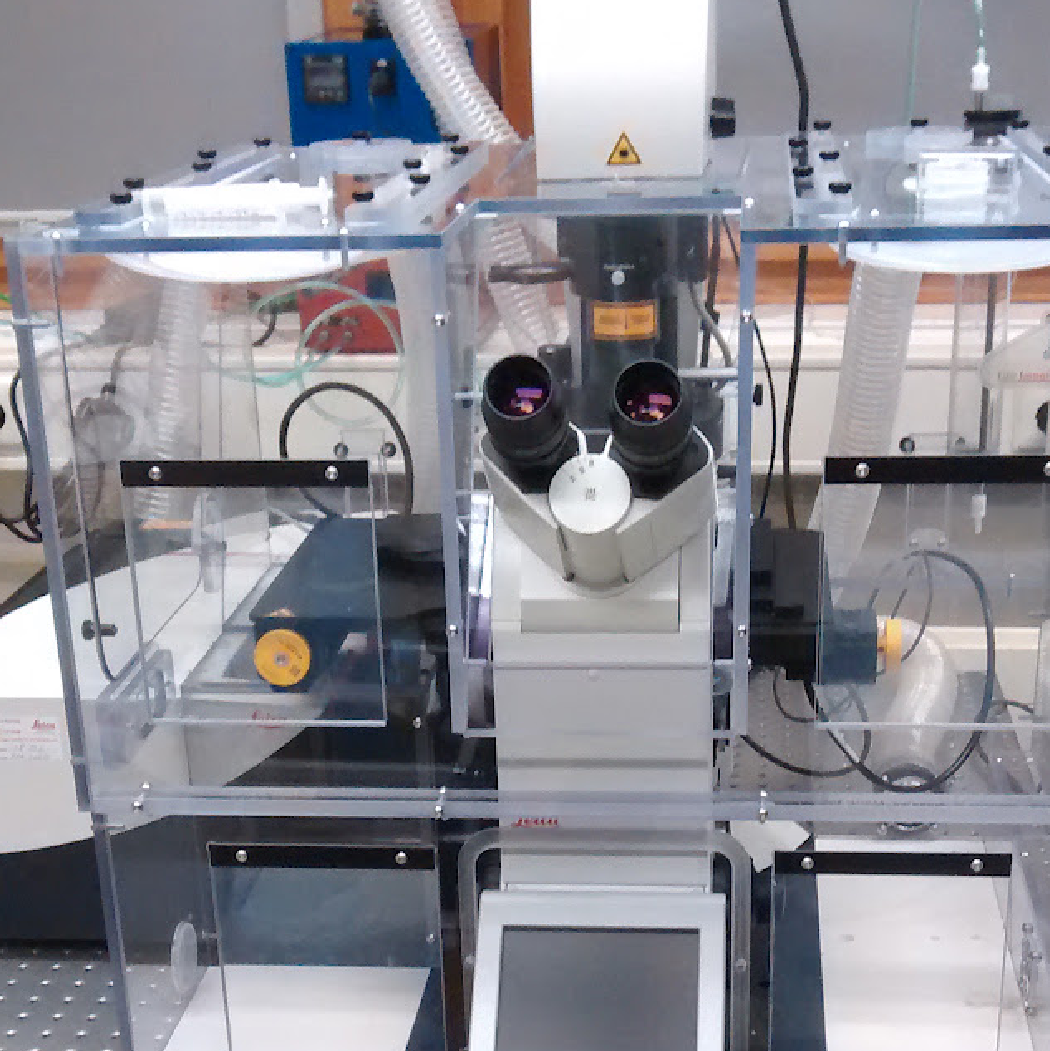
Inverted DMi8 microscope with a lightsheet unit; Ex lasers: 405-nm, 488-nm; 561-nm; 633-nm; Hamamatsu Flash 4.0 V3 sCMOS camera; L10x/0.30 W DLS; L25x/0.95 W DLS; 10x/0.40; 20x/0.75 IMM; 40x/1.10 W; 63x/1.30 Glyc.
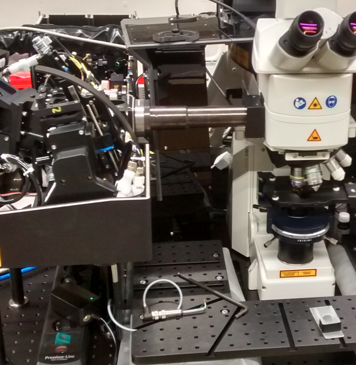
2-photon laser (690-1040 nm); Upright microscope; 16x/0.6 IMM CORR VISIR, 20x/0.5 W, 40x/0.80 W, 63x/0.9 W; 4-Tune 3-channels (2x HyDs); 2x HyDs for internal detection; FALCON (FLIM).
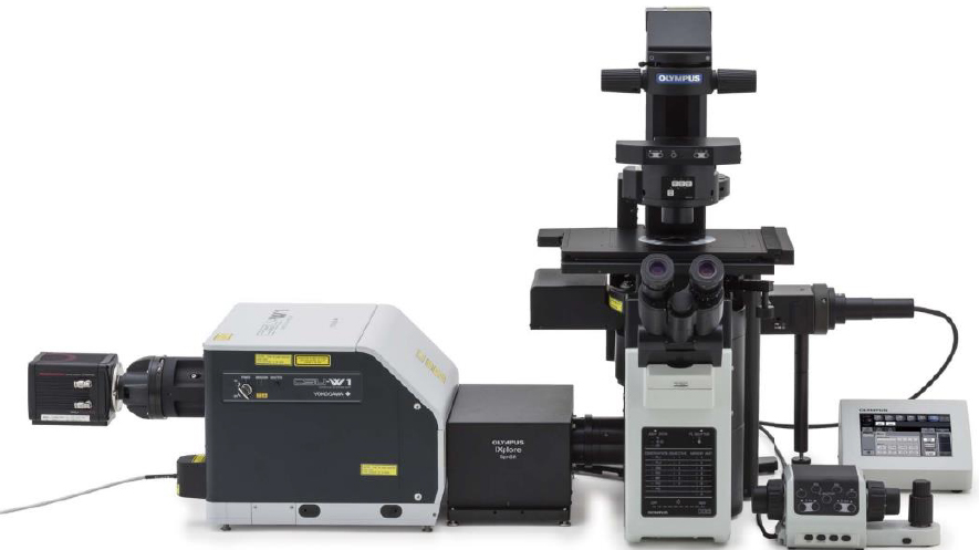
4 lasers (405, 488, 561, 640-nm), Yokogawa W1 spinning disc, 2x Hamamatsu Orca Fusion, Piezo z-stage, temp. and CO2 incubator, water dispenser, software CellSense + ScanR, 4x/0.16 dry, 10x/0.40 dry, 20x/0.80 dry, 20x/0.70 water, 30x/1.05 silicone, 40x/0.95 dry, 60x/1.50 oil, 60x/1.20 water, 100x/1.35 silicone
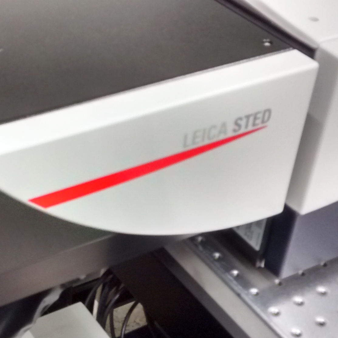
Inverted microscope with a white light laser. Located in the basement of building 439. White light laser + 405 nm DMOD, 2x HyD, 20x/0.75 IMM, 40x/1.10 W, 93x/1.30 Glyc motC, STED 3X 775-nm; FALCON (FLIM).
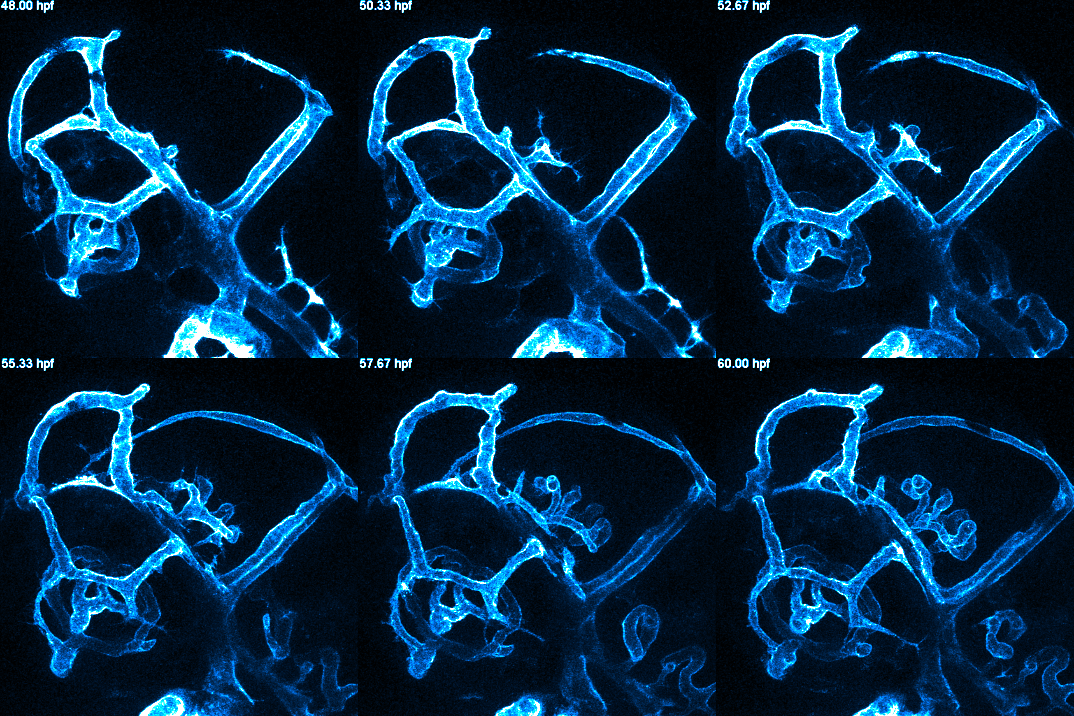
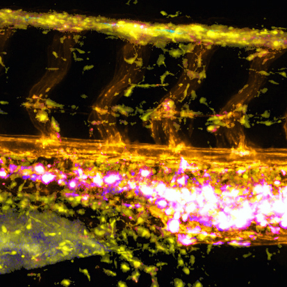
Inverted DMi8 microscope with environment controls
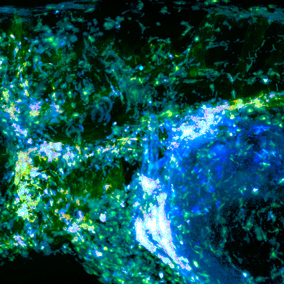
Upright DM6 microscope
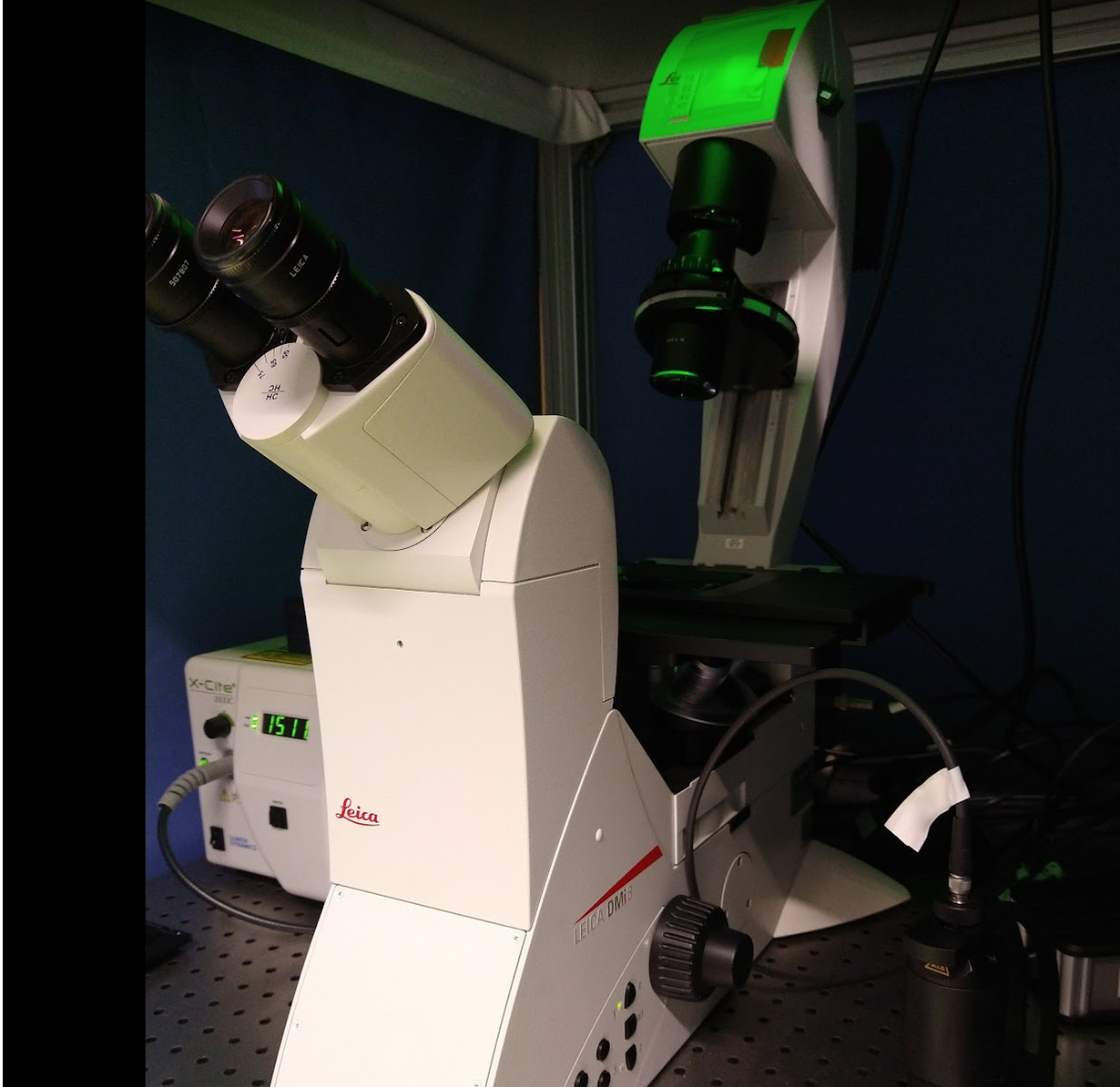
with colour camera
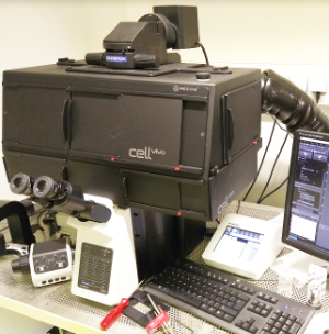
PRIMO (micro-patterning) & luminescence imaging
Compound fluorescence microscopy with motorized XYZ stage
high throughput cell culture screen

operation voltage at 80 kV