
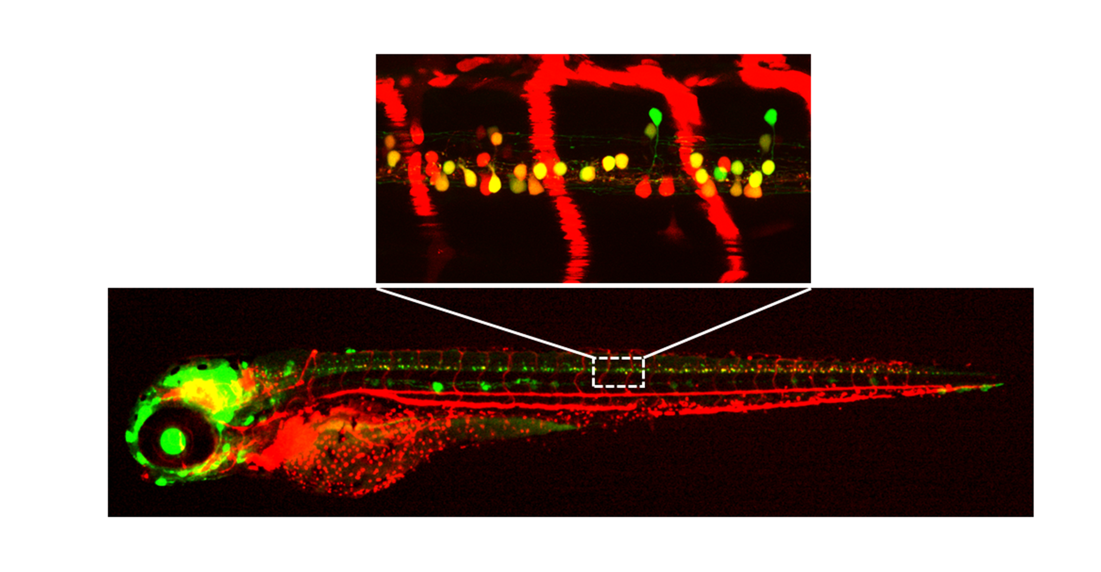
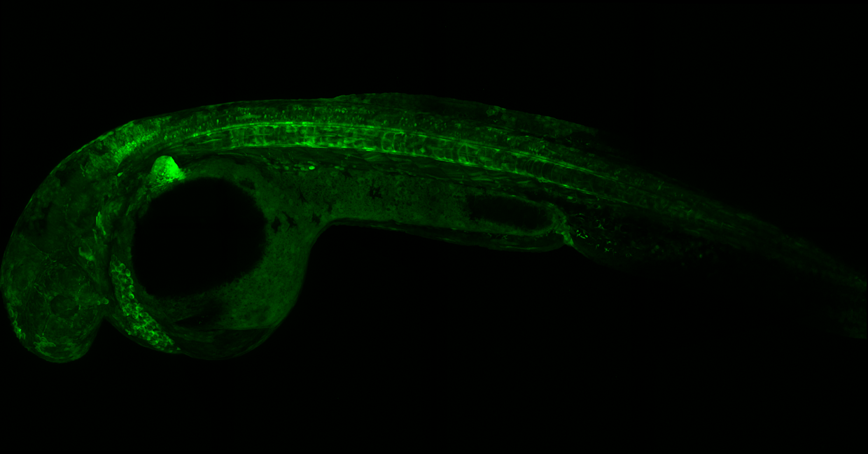
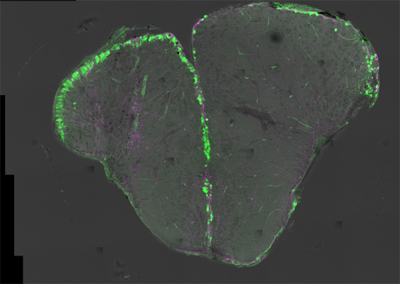
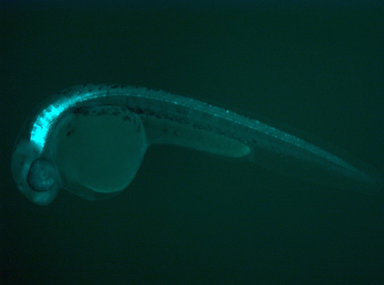
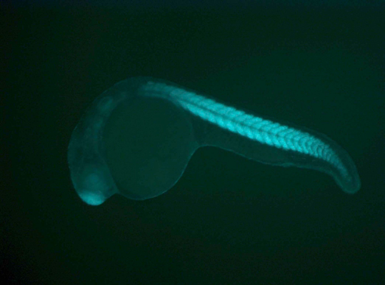
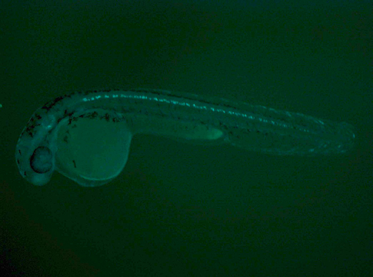
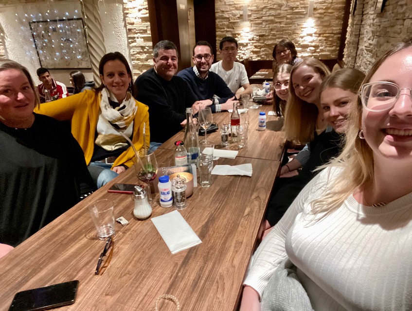
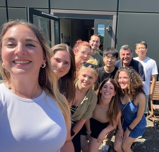
Zebrafish Neurodevelopment & Regeneration
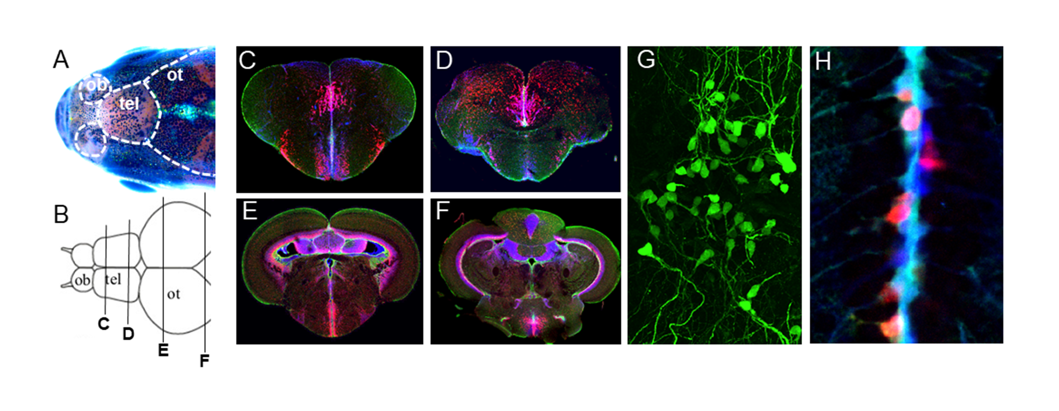
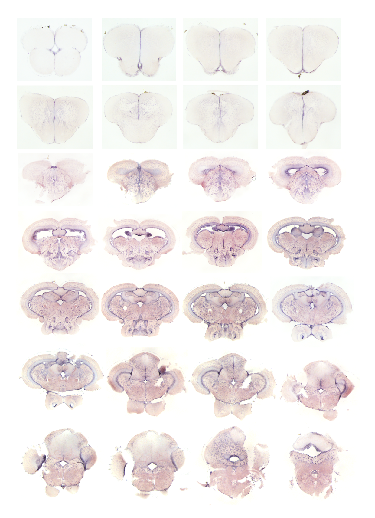
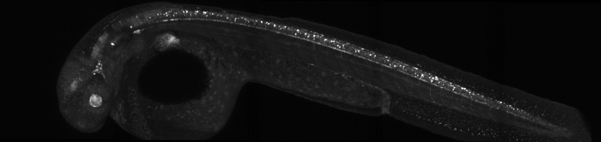
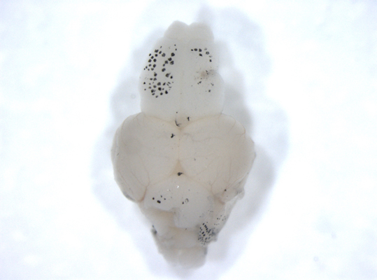
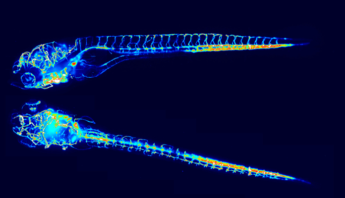
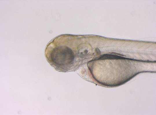
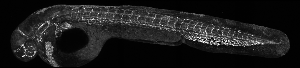
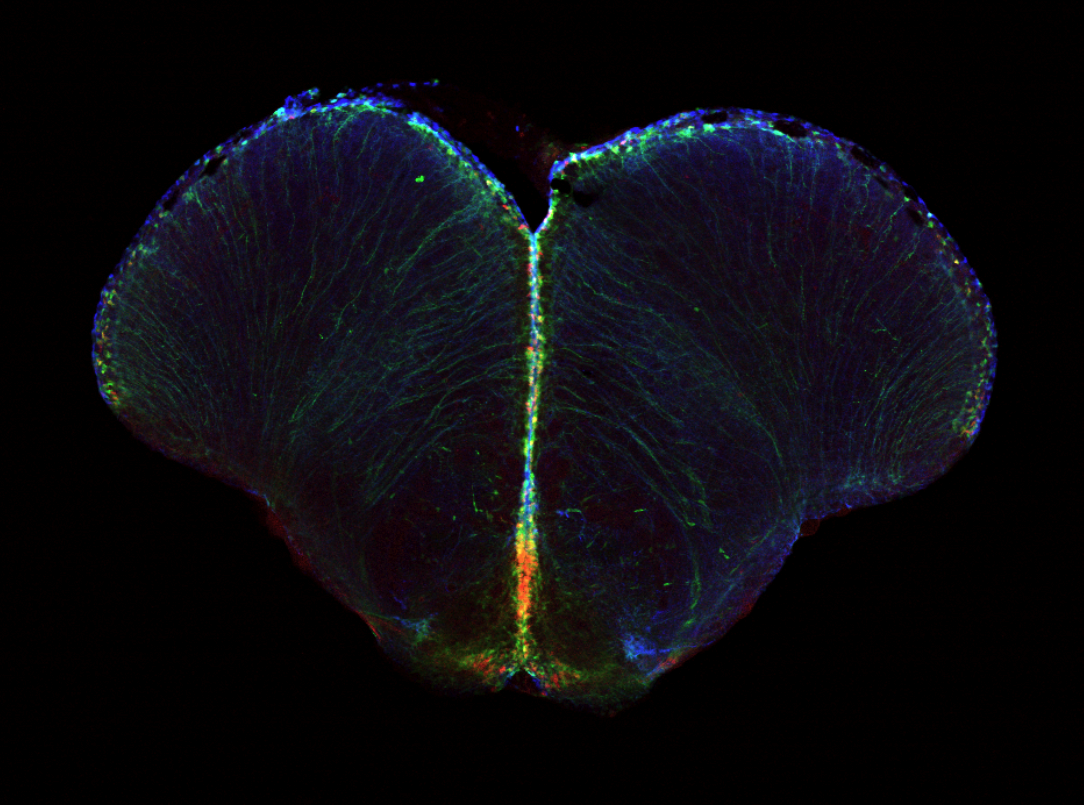
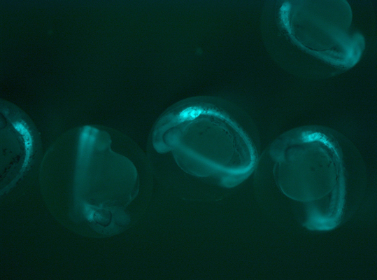
We study how the nervous system develops, functions, and regenerates—focusing on how diverse types of neurons are generated and maintained. Our lab uses zebrafish as a primary model due to its genetic similarity to humans, transparency, and exceptional regenerative abilities. Our research centres on neurogenesis and regeneration, using the embryonic spinal cord and adult brain to explore how neurons form, differentiate, and recover after injury. We aim to decode the complex networks of transcriptional regulators (TRs) and cis-regulatory elements (CREs) that control gene expression during these processes. To do this, we apply cutting-edge techniques like CRISPR/Cas9 gene editing, transgenic reporters, single-cell sequencing, and high-throughput genomics (e.g., RNA-seq, CAGE-seq, ATAC-seq). We also collaborate with chemists to identify natural and synthetic compounds that influence key signalling pathways (BMP, Notch, Shh) and explore molecules with photomodulatory activity for neuroregeneration. Ultimately, our goal is to uncover fundamental mechanisms of neuronal development and repair—laying the groundwork for future regenerative therapies and engineered neuronal tissues.
Sepand Rastegar
- Group: Rastegar
- ORCID
Karlsruhe Institute of Technology (KIT)
Campus North
Institute of Biological and Chemical Systems (IBCS)
Building 319
Hermann-von-Helmholtz-Platz 1
76344 Eggenstein-Leopoldshafen
Germany
Publications
Chen, J.; Takamiya, M.; Hendriks, A.; Beil, T.; Várnai, C.; Diotel, N.; Rastegar, S.
2026. The FEBS Journal, 293 (7), 1951–1969. doi:10.1111/febs.70345
Pansera, L.; Pagliari, S.; Mhalhel, K.; Aragona, M.; Sicari, M.; Galeano, M.; Colonna, M. R.; Levanti, M.; Laurà, R.; Abbate, F.; Cicero, N.; Labra, M.; Rastegar, S.; Germanà, A.; Campone, L.; Montalbano, G.
2026. International Journal of Molecular Sciences, 27 (4), Art.-Nr.: 1687. doi:10.3390/ijms27041687
Chen, J.
2025, March 13. Karlsruher Institut für Technologie (KIT). doi:10.5445/IR/1000179272
Yan, J.; Takamiya, M.; Zhang, D.; Pace, G.; Rastegar, S.; Wang, H.; Schoch, S.; Köberle, B.; Hartwig, A.; Dickmeis, T.; Weiss, C.
2025. Environment International, 197, Article no: 109349. doi:10.1016/j.envint.2025.109349
Gence, L.; Morello, E.; Rastegar, S.; Apalama, M. L.; Meilhac, O.; Bascands, J.-L.; Diotel, N.
2024. European Journal of Neuroscience. doi:10.1111/ejn.16586
Chen, J.; Sanchez-Iranzo, H.; Diotel, N.; Rastegar, S.
2024. The FEBS Journal, 291 (19), 4193–4205. doi:10.1111/febs.17231
Cucun, G.
2024, August 28. Karlsruher Institut für Technologie (KIT). doi:10.5445/IR/1000173043
Chen, F.
2024, August 27. Karlsruher Institut für Technologie (KIT). doi:10.5445/IR/1000173042
Cucun, G.; Köhler, M.; Pfitsch, S.; Rastegar, S.
2024. The FEBS Journal, 291 (4), 646–662. doi:10.1111/febs.16913
Seliwjorstow, A.; Takamiya, M.; Rastegar, S.; Pianowski, Z.
2024. ChemBioChem, 25 (8), Article e202400143. doi:10.1002/cbic.202400143
Štefl, M.; Takamiya, M.; Middel, V.; Tekpınar, M.; Nienhaus, K.; Beil, T.; Rastegar, S.; Strähle, U.; Nienhaus, G. U.
2024. iScience, 27 (2), 108849. doi:10.1016/j.isci.2024.108849
Fernezelian, D.; Pfitsch, S.; Rastegar, S.; Diotel, N.
2024. Neural Development, 19, Article no: 17. doi:10.1186/s13064-024-00195-1
Pagliari, S.; Sicari, M.; Pansera, L.; Guidi Nissim, W.; Mhalhel, K.; Rastegar, S.; Germanà, A.; Cicero, N.; Labra, M.; Cannavacciuolo, C.; Montalbano, G.; Campone, L.
2024. Journal of Food Science, 89 (6), 3729–3744. doi:10.1111/1750-3841.17079
Pellegrini, E.; Fernezelian, D.; Malleret, C.; Gueguen, M. M.; Patche-Firmin, J.; Rastegar, S.; Meilhac, O.; Diotel, N.
2023. The journal of comparative neurology, 531 (17), 1828–1845. doi:10.1002/cne.25543
Chen, F.; Köhler, M.; Cucun, G.; Takamiya, M.; Kizil, C.; Cosacak, M. I.; Rastegar, S.
2023. iScience, 26 (8), Artkl. Nr.: 107342. doi:10.1016/j.isci.2023.107342
Narra, S. S.; Rondeau, P.; Fernezelian, D.; Gence, L.; Ghaddar, B.; Bourdon, E.; Lefebvre d’Hellencourt, C.; Rastegar, S.; Diotel, N.
2023. Journal of Comparative Neurology, 531 (2), 238–255. doi:10.1002/cne.25421
Gence, L.; Fernezelian, D.; Meilhac, O.; Rastegar, S.; Bascands, J.-L.; Diotel, N.
2023. Journal of Comparative Neurology, 531 (17), 1812–1827. doi:10.1002/cne.25542
Hoffmann, M.; Gerlach, S.; Takamiya, M.; Tarazi, S.; Hersch, N.; Csiszár, A.; Springer, R.; Dreissen, G.; Scharr, H.; Rastegar, S.; Beil, T.; Strähle, U.; Merkel, R.; Hoffmann, B.
2023. Pharmaceutics, 15 (4), Art.-Nr.: 1210. doi:10.3390/pharmaceutics15041210
Mhalhel, K.; Sicari, M.; Chen, J.; Levanti, M.; Diotel, N.; Pansera, L.; Rastegar, S.; Germanà, A.; Montalbano, G.
2023. Cells, 12 (2), Art.-Nr.: 252. doi:10.3390/cells12020252
Punampalam, R.
2022, November 28. Karlsruher Institut für Technologie (KIT). doi:10.5445/IR/1000152185
Khan, A.; Molitor, A.; Mayeur, S.; Zhang, G.; Rinaldi, B.; Lannes, B.; Lhermitte, B.; Umair, M.; Arold, S. T.; Friant, S.; Rastegar, S.; Anheim, M.; Bahram, S.; Carapito, R.
2022. Movement disorders, 37 (2), 365–374. doi:10.1002/mds.28861
Hansjosten, I.; Takamiya, M.; Rapp, J.; Reiner, L.; Fritsch-Decker, S.; Mattern, D.; Andraschko, S.; Anders, C.; Pace, G.; Dickmeis, T.; Peravali, R.; Rastegar, S.; Strähle, U.; Hsiao, I.-L.; Gilliland, D.; Ojea-Jimenez, I.; Ambrose, S. V. Y.; Belinga-Desaunay-Nault, M.-F. A.; Khan, A. O.; Lynch, I.; Valsami-Jones, E.; Diabaté, S.; Weiss, C.
2022. Environmental science / Nano, 91 (1), 375–392. doi:10.1039/d1en00299f
Wesseler, F.; Lohmann, S.; Riege, D.; Halver, J.; Roth, A.; Pichlo, C.; Weber, S.; Takamiya, M.; Müller, E.; Ketzel, J.; Flegel, J.; Gihring, A.; Rastegar, S.; Bertrand, J.; Baumann, U.; Knippschild, U.; Peifer, C.; Sievers, S.; Waldmann, H.; Schade, D.
2022. Journal of Medicinal Chemistry, 65 (22), 15263–15281. doi:10.1021/acs.jmedchem.2c01199
Baranasic, D.; Hörtenhuber, M.; Balwierz, P. J.; Zehnder, T.; Mukarram, A. K.; Nepal, C.; Várnai, C.; Hadzhiev, Y.; Jimenez-Gonzalez, A.; Li, N.; Wragg, J.; D’Orazio, F. M.; Relic, D.; Pachkov, M.; Díaz, N.; Hernández-Rodríguez, B.; Chen, Z.; Stoiber, M.; Dong, M.; Stevens, I.; Ross, S. E.; Eagle, A.; Martin, R.; Obasaju, O.; Rastegar, S.; McGarvey, A. C.; Kopp, W.; Chambers, E.; Wang, D.; Kim, H. R.; Acemel, R. D.; Naranjo, S.; Łapiński, M.; Chong, V.; Mathavan, S.; Peers, B.; Sauka-Spengler, T.; Vingron, M.; Carninci, P.; Ohler, U.; Lacadie, S. A.; Burgess, S. M.; Winata, C.; van Eeden, F.; Vaquerizas, J. M.; Gómez-Skarmeta, J. L.; Onichtchouk, D.; Brown, B. J.; Bogdanovic, O.; van Nimwegen, E.; et al.
2022. Nature Genetics, 54, 1037–1050. doi:10.1038/s41588-022-01089-w
Lübke, L.; Zhang, G.; Strähle, U.; Rastegar, S.
2022. Brain Sciences, 12 (2), Art.Nr. 284. doi:10.3390/brainsci12020284
Hüpfel, M.; Fernández Merino, M.; Bennemann, J.; Takamiya, M.; Rastegar, S.; Tursch, A.; Holstein, T. W.; Nienhaus, G. U.
2022. Biomedical optics express, 13 (1), 147–158. doi:10.1364/BOE.443660
Sulliman, N. C.; Ghaddar, B.; Gence, L.; Patche, J.; Rastegar, S.; Meilhac, O.; Diotel, N.
2021. Scientific reports, 11 (1), Art. Nr.: 6439. doi:10.1038/s41598-021-85183-9
Zhang, G.; Lübke, L.; Chen, F.; Beil, T.; Takamiya, M.; Diotel, N.; Strähle, U.; Rastegar, S.
2021. Cells, 10 (10), Art.-Nr.: 2794. doi:10.3390/cells10102794
Afonin, S.; Koniev, S.; Préau, L.; Takamiya, M.; Strizhak, A. V.; Babii, O.; Hrebonkin, A.; Pivovarenko, V. G.; Dathe, M.; Noble, F. le; Rastegar, S.; Strähle, U.; Ulrich, A. S.; Komarov, I. V.
2021. Frontiers in Chemistry, 9, Art. Nr.: 688446. doi:10.3389/fchem.2021.688446
Gourain, V.; Armant, O.; Lübke, L.; Diotel, N.; Rastegar, S.; Strähle, U.
2021. Frontiers in neuroscience, 15, Article no: 671249. doi:10.3389/fnins.2021.671249
Ghaddar, B.; Lübke, L.; Couret, D.; Rastegar, S.; Diotel, N.
2021. Cells, 10 (2), 391. doi:10.3390/cells10020391
Diotel, N.; Lübke, L.; Strähle, U.; Rastegar, S.
2020. Frontiers in neuroscience, 14, Art.-Nr.: 568930. doi:10.3389/fnins.2020.568930
Takamiya, M.; Stegmaier, J.; Kobitski, A. Y.; Schott, B.; Weger, B. D.; Margariti, D.; Cereceda Delgado, A. R.; Gourain, V.; Scherr, T.; Yang, L.; Sorge, S.; Otte, J. C.; Hartmann, V.; Wezel, J. van; Stotzka, R.; Reinhard, T.; Schlunck, G.; Dickmeis, T.; Rastegar, S.; Mikut, R.; Nienhaus, G. U.; Strähle, U.
2020. PLoS Genetics, 16 (6), Art. Nr.: e1008774. doi:10.1371/journal.pgen.1008774
Zhang, G.; Ferg, M.; Lübke, L.; Takamiya, M.; Beil, T.; Gourain, V.; Diotel, N.; Strähle, U.; Rastegar, S.
2020. Stem cells, 38, 875–889. doi:10.1002/stem.3182
Baanannou, A.; Rastegar, S.; Bouzid, A.; Takamiya, M.; Gerber, V.; Souissi, A.; Beil, T.; Jrad, O.; Strähle, U.; Masmoudi, S.
2020. Development genes and evolution, 230 (1), 37. doi:10.1007/s00427-020-00647-8
Baanannou, A.; Rastegar, S.; Bouzid, A.; Takamiya, M.; Gerber, V.; Souissi, A.; Beil, T.; Jrad, O.; Strähle, U.; Masmoudi, S.
2020. Development genes and evolution, 230, 27–36. doi:10.1007/s00427-019-00642-8
Rastegar, S.; Parimisetty, A.; Cassam Sulliman, N.; Narra, S. S.; Weber, S.; Rastegar, M.; Viranaicken, W.; Couret, D.; Planesse, C.; Strähle, U.; Meilhac, O.; Lefebvre d’Hellencourt, C.; Diotel, N.
2019. The journal of comparative neurology, 527 (14), 2317–2333. doi:10.1002/cne.24669
Gerber, V.; Yang, L.; Takamiya, M.; Ribes, V.; Gourain, V.; Peravali, R.; Stegmaier, J.; Mikut, R.; Reischl, M.; Ferg, M.; Rastegar, S.; Strähle, U.
2019. Development <Cambridge>, 146 (4), dev172510. doi:10.1242/dev.172510
Weger, M.; Diotel, N.; Weger, B. D.; Beil, T.; Zaucker, A.; Eachus, H. L.; Oakes, J. A.; Rego, J. L. do; Storbeck, K.-H.; Gut, P.; Strähle, U.; Rastegar, S.; Müller, F.; Krone, N.
2018. Journal of neuroendocrinology, 30 (4), Art.Nr.: e12586. doi:10.1111/jne.12586
Angelin, A.; Kassel, O.; Rastegar, S.; Strähle, U.; Niemeyer, C. M.
2017. ChemistryOpen, 6 (1), 33–39. doi:10.1002/open.201600153
Middel, V.; Zhou, L.; Takamiya, M.; Beil, T.; Shahid, M.; Roostalu, U.; Grabher, C.; Rastegar, S.; Reischl, M.; Nienhaus, G. U.; Strähle, U.
2016. Nature Communications, 7, 12875. doi:10.1038/ncomms12875
Rastegar, S.; Strähle, U.
2016. Genetics, Genomics and Phenomics of Fish. Ed.: N.S. Foulkes, 195–216, Academic Press. doi:10.1016/bs.adgen.2016.04.003
Strähle, U.; Ferg, M.; Armant, O.; Gradl, M.; Kaufmann, L.; Rastegar, S.
2016. Zebrafish, 13 (3), 234–235. doi:10.1089/zeb.2016.29003.str
Takamiya, M.; Xu, F.; Suhonen, H.; Gourain, V.; Yang, L.; Yu Ho, N.; Helfen, L.; Schröck, A.; Etard, C.; Grabher, C.; Rastegar, S.; Schlunck, G.; Reinhard, T.; Baumbach, T.; Strähle, U.
2016. Scientific reports, 6, Art. Nr.: 25046. doi:10.1038/srep25046
Shahid, M.; Takamiya, M.; Stegmaier, J.; Middel, V.; Gradl, M.; Klüver, N.; Mikut, R.; Dickmeis, T.; Scholz, S.; Rastegar, S.; Yang, L.; Strähle, U.
2016. Scientific Reports, 6, Art.Nr. 23768. doi:10.1038/srep23768
Diotel, N.; Beil, T.; Strähle, U.; Rastegar, S.
2015. Gene Expression Patterns, 19 (1-2), 1–13. doi:10.1016/j.gep.2015.05.004
Naville, M.; Ishibashi, M.; Ferg, M.; Bengani, H.; Rinkwitz, S.; Krecsmarik, M.; Hawkins, T. A.; Wilson, S. W.; Manning, E.; Chilamakuri, C. S. R.; Wilson, D. I.; Louis, A.; Raymond, F. L.; Rastegar, S.; Strähle, U.; Lenhard, B.; Bally-Cuif, L.; Heyningen, V. van; FitzPatrick, D. R.; Becker, T. S.; Crollius, H. R.
2015. Nature Communications, 6, 6904. doi:10.1038/ncomms7904
Rodriguez Viales, R.; Diotel, N.; Ferg, M.; Armant, O.; Eich, J.; Alunni, A.; März, M.; Bally-Cuif, L.; Rastegar, S.; Strähle, U.
2015. Stem cells, 33, 892–903. doi:10.1002/stem.1883
Diotel, N.; Rodriguez Vialez, R.; Armant, O.; März, M.; Ferg, M.; Rastegar, S.; Strähle, U.
2015. The journal of comparative neurology, 523 (8), 1202–1221. doi:10.1002/cne.23733
Takamiya, M.; Weger, B. D.; Schindler, S.; Beil, T.; Yang, L.; Armant, O.; Ferg, M.; Schlunck, G.; Reinhard, T.; Dickmeis, T.; Rastegar, S.; Strähle, U.; Leung, Y. F.
2015. PLoS ONE, 10 (2), e0117645. doi:10.1371/journal.pone.0117645
Kobitski, A. Y.; Otte, J. C.; Takamiya, M.; Schäfer, B.; Mertes, J.; Stegmaier, J.; Rastegar, S.; Rindone, F.; Hartmann, V.; Stotzka, R.; Garcia, A.; Wezel, J. van; Mikut, R.; Strähle, U.; Nienhaus, G. U.
2015. Scientific Reports, 5 (8601), 1–10. doi:10.1038/srep08601
Schmidt, R.; Beil, T.; Strähle, U.; Rastegar, S.
2014. Journal of Visualized Experiments, 90, e51753/1–8. doi:10.3791/51753
Ferg, M.; Rastegar, S.; Strähle, U.
2014. Biospektrum, 2, 148–150. doi:10.1007/s12268-014-0419-0
Ferg, M.; Armant, O.; Yang, L.; Dickmeis, T.; Rastegar, S.; Strähle, U.
2014. Briefings in functional genomics, 13, 131–143. doi:10.1093/bfgp/elt044
Stegmaier, J.; Shahid, M.; Takamiya, M.; Yang, L.; Rastegar, S.; Reischl, M.; Strähle, U.; Mikut, R.
2014. Bioinformatics, 30, 726–733. doi:10.1093/bioinformatics/btt600
Nepal, C.; Hadzhiev, Y.; Previti, C.; Haberle, V.; Li, N.; Takahashi, H.; Suzuki, A. M.; Sheng, Y.; Abdelhamid, R. F.; Anand, S.; Gehrig, J.; Akalin, A.; Kockx, C. E. M.; Sloot, A. A. J. van der; Ijcken, W. F. J. van; Armant, O.; Rastegar, S.; Watson, C.; Strähle, U.; Stupka, E.; Carninci, P.; Lenhard, B.; Müller, F.
2013. Genome Research, 23, 1938–1950. doi:10.1101/gr.153692.112
Armant, O.; März, M.; Schmidt, R.; Ferg, M.; Diotel, N.; Ertzer, R.; Bryne, J. C.; Yang, L.; Baader, I.; Reischl, M.; Legradi, J.; Mikut, R.; Stemple, D.; Ilcken, W. van; Sloot, A. van der; Lenhard, B.; Strähle, U.; Rastegar, S.
2013. Developmental Biology, 380, 351–362. doi:10.1016/j.ydbio.2013.05.006
Weger, M.; Weger, B. D.; Diotel, N.; Rastegar, S.; Hirota, T.; Kay, S. A.; Strähle, U.; Dickmeis, T.
2013. Developmental Biology, 380, 259–273. doi:10.1016/j.ydbio.2013.04.035
Ho, N. Y.; Yang, L.; Legradi, J.; Armant, O.; Takamiya, M.; Rastegar, S.; Strähle, U.
2013. Environmental Science and Technology, 47, 3316–3325. doi:10.1021/es3050967
Strähle, U.; Scholz, S.; Geisler, R.; Greiner, P.; Hollert, H.; Rastegar, S.; Schumacher, A.; Selderslaghs, I.; Weiss, C.; Witters, H.; Braunbeck, T.
2012. Reproductive toxicology, 33, 128–132. doi:10.1016/j.reprotox.2011.06.121
März, M.; Schmidt, R.; Rastegar, S.; Strähle, U.
2011. Developmental Dynamics, 240, 2221–2231. doi:10.1002/dvdy.22710
März, M.; Schmidt, R.; Rastegar, S.; Strähle, U.
2010. Developmental Dynamics, 239, 3336–3349. doi:10.1002/dvdy.22455
Keiter, S.; Peddinghaus, S.; Feiler, U.; Goltz, B. von der; Hafner, C.; Ho, N. Y.; Rastegar, S.; Otte, J. C.; Ottermanns, R.; Reifferscheid, G.; Strähle, U.; Braunbeck, T.; Hammers-Wirtz, M.; Hollert, H.
2010. Umweltwissenschaften und Schadstoff-Forschung, 22, 94–98. doi:10.1007/s12302-010-0119-4
Yang, L.; Rastegar, S.; Strähle, U.
2010. Development, 137, 2713–22. doi:10.1242/dev.048470
Keiter, S.; Peddinghaus, S.; Feiler, U.; Goltz, B. von der; Hafner, C.; Ho, N. Y.; Rastegar, S.; Otte, J. C.; Ottermanns, R.; Reifferscheid, G.; Strähle, U.; Braunbeck, T.; Hammers-Wirtz, M.; Hollert, H.
2010. Journal of Soils and Sediments, 10, 714–17. doi:10.1007/s11368-010-0221-7
Rastegar, S.; Hess, I.; Dickmeis, T.; Nicod, J. C.; Ertzer, R.; Hadzhiev, Y.; Thies, W. G.; Scherer, G.; Strähle, U.
2008. Developmental Biology, 318, 366–77. doi:10.1016/j.ydbio.2008.03.034
Strähle, U.; Rastegar, S.
2008. Brain Research Bulletin, 75, 225–30. doi:10.1016/j.brainresbull.2007.11.010
Vinothkumar, S.; Rastegar, S.; Takamiya, M.; Ertzer, R.; Strähle, U.
2008. Developmental Biology, 314, 200–214. doi:10.1016/j.ydbio.2007.11.034
Strähle, U.; März, M.; Rathnam, S.; Rastegar, S.
2007. Nachrichten - Forschungszentrum Karlsruhe, 39, 133–39
Ertzer, R.; Müller, F.; Hadzhiev, Y.; Rathnam, S.; Fischer, N.; Rastegar, S.; Strähle, U.
2007. Developmental Biology, 301, 578–89. doi:10.1016/j.ydbio.2006.11.004
Strähle, U.; Rastegar, S.; Etard, C.; Weg-Remers, S.; Müller, F.
2006. Nachrichten - Forschungszentrum Karlsruhe, 38, 93–95
Lam, C. S.; Rastegar, S.; Strähle, U.
2006. Neuroscience, 138, 83–95. doi:10.1016/j.neuroscience.2005.10.069
Norton, W. H.; Mangoli, M.; Lele, Z.; Pogoda, H. M.; Diamond, B.; Mercurio, S.; Russell, C.; Teraoka, H.; Stickney, H. L.; Rauch, G. J.; Heisenberg, C. P.; Houart, C.; Schilling, T. F.; Frohnhoefer, H. G.; Rastegar, S.; Neumann, C. J.; Gardiner, R. M.; Strähle, U.; Geisler, R.; Rees, M.; Talbot, W. S.; Wilson, S. W.
2005. Development, 132, 645–58
Blader, P.; Lam, C. S.; Rastegar, S.; Scardigli, R.; Nicod, J. C.; Simplicio, N.; Plessy, C.; Fischer, N.; Schuurmans, C.; Guillemot, F.; Strähle, U.
2004. Development, 131, 5627–37. doi:10.1242/dev.01455
Geling, A.; Plessy, C.; Rastegar, S.; Strähle, U.; Bally-Cuif, L.
2004. Development, 131, 1993–2006
Strähle, U.; Lam, C. S.; Ertzer, R.; Rastegar, S.
2004. Trends in Genetics, 20, 155–62. doi:10.1016/j.tig.2004.01.002
Henningfeld, K. A.; Friedle, H.; Rastegar, S.; Knöchel, W.
2002. Journal of Biological Chemistry, 277 (3), 2097–2103. doi:10.1074/jbc.M108524200
Rastegar, S.; Albert, S.; Le Roux, I.; Fischer, N.; Blader, P.; Müller, F.; Strähle, U.
2002. Developmental biology, (2002), 252 (1), 1–14. doi:10.1006/dbio.2002.0837
Henningfeld, K. A.; Rastegar, S.; Adler, G.; Knöchel, W.
2000. Journal of Biological Chemistry, 275 (29), 21827–21835. doi:10.1074/jbc.M000978200
Rastegar, S.
1999. Mechanisms of development, 81 ((1-2), 139–149. doi:10.1016/s0925-4773(98)00239-1
Blader, P.; Rastegar, S.; Fischer, N.; Strähle, U.
1997. Science, 278 (5345), 1937–1940. doi:10.1126/science.278.5345.1937